
Does a Suspicious Mole Mean Skin Cancer?
No, a suspicious mole does not necessarily mean skin cancer. It means your GP has identified features that require further assessment. Changes in size, shape, colour or appearance may raise concern, but these features alone cannot confirm a diagnosis.
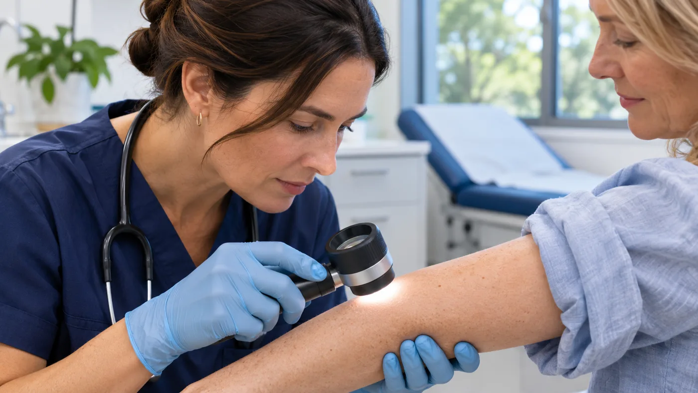
During an examination, your GP may use a dermatoscope, a handheld magnifying instrument that provides a closer view of the lesion. According to Cancer Council Australia, harmless skin spots and skin cancers can look similar, so a biopsy may be needed when the diagnosis remains uncertain.
A biopsy may identify:
- Benign lesions: Non-cancerous moles or harmless skin growths.
- Atypical or precancerous changes: Abnormal tissue that may require monitoring or treatment.
- Skin cancer: Conditions such as basal cell carcinoma, squamous cell carcinoma or melanoma.
Changes in a mole’s appearance are a reason for getting a mole checked by a GP, but the recommendation for a biopsy means the diagnosis still needs clarification.
Why Might a GP Recommend a Biopsy or Excision?
A GP may recommend a biopsy or excision when a suspicious skin lesion requires further investigation to confirm a diagnosis or guide treatment. During a skin cancer check, your doctor assesses the lesion’s appearance, changes over time, location and relevant medical history before deciding whether tissue examination is necessary.
A biopsy or excision may be recommended when:
- A mole has features that raise concern for melanoma or another skin cancer.
- A lesion has changed significantly, is bleeding or has developed an ulcer that is not healing.
- The diagnosis remains uncertain after a clinical examination.
- A tissue sample is needed to confirm the diagnosis and plan appropriate treatment.
Not every unusual skin lesion needs removal. Some can be monitored, while others require prompt investigation. The Cancer Council’s melanoma guide recommends biopsy within two weeks of the initial GP consultation when melanoma is suspected. Your GP may perform the procedure or refer you to a specialist, depending on the lesion and clinical expertise.
What Is the Difference Between a Biopsy and an Excision?
A biopsy involves removing tissue for laboratory examination, while an excision involves surgically removing a skin lesion. The procedures can overlap, as removing an entire suspicious mole for pathology is called an excisional biopsy. The method depends on the lesion’s characteristics and suspected diagnosis.
| Procedure | What Happens | When It May Be Used |
| Excisional biopsy | Removes the entire lesion with a small margin of surrounding skin. | Generally preferred when melanoma is suspected. |
| Punch biopsy | Uses a circular instrument to remove a cylindrical skin sample. | When sampling part of a lesion is clinically appropriate. |
| Shave biopsy | Removes a superficial portion of a skin lesion using a blade. | For selected superficial lesions. |
| Surgical excision | Removes a lesion, sometimes with a wider margin of surrounding tissue. | For treatment or further removal following pathology. |
Why Is Excisional Biopsy Preferred for Suspected Melanoma?
An excisional biopsy is generally preferred when melanoma is suspected because removing the entire lesion allows the pathologist to assess its structure and measure tumour thickness, which helps guide treatment decisions.
A partial biopsy may be considered when complete removal is difficult due to the lesion’s size or location. However, examining only part of a lesion can limit the information available, making careful selection of the biopsy technique important.
What Happens During a Mole Biopsy?
A mole biopsy usually involves numbing the skin with local anaesthetic, removing the required tissue and dressing or closing the wound. The sample is then sent to a pathology laboratory for examination. The procedure varies depending on the biopsy method and the lesion’s location.
Step 1: Preparing for the Procedure
Before the biopsy, your clinician explains the recommended technique, what to expect and possible risks, including bleeding, infection and scarring. You can also ask whether stitches will be needed and what recovery may involve.
Inform your clinician about any allergies, medical conditions or medicines you take, particularly blood thinners. Do not stop prescribed medication unless your treating clinician advises you to do so.
Step 2: Numbing and Removing the Tissue
The clinician cleans the area and injects a local anaesthetic to numb the skin. You may feel a brief sting from the injection or pressure during the procedure, but tell your clinician if you experience pain.
Once the area is numb, the clinician removes part or all of the suspicious lesion using the selected biopsy technique. The amount of tissue removed depends on the suspected diagnosis and the purpose of the procedure.
Step 3: Closing and Dressing the Wound
After removing the tissue, the clinician controls any bleeding and closes the wound if necessary. Some biopsies require stitches, while smaller or more superficial wounds may heal without them.
A dressing may be applied, and your clinician will provide instructions about wound care, activity restrictions and follow-up appointments. The procedure may leave a scar, depending on the wound’s size, location and healing process.
Step 4: Sending the Sample for Pathology
The sample is labelled and sent for pathology. Before leaving, ask when and how you will receive the results.
What Happens to the Sample After the Biopsy?
At the pathology laboratory, a pathologist examines thin sections of the tissue under a microscope to identify the lesion and assess abnormal or cancerous cells.
The report may describe the lesion type, relevant cell features and whether abnormal tissue reaches the sample’s edges, known as margins. For melanoma, it may also record Breslow thickness, which measures tumour depth and helps guide treatment.
Your GP considers the report alongside the clinical examination. If the findings do not explain the suspicious lesion, further examination or sampling may be needed.
How Long Do Mole Biopsy Results Take?
Mole biopsy results often take around one to two weeks, but timing varies by laboratory and the tests required. Ask the treating practice for its expected reporting timeframe.
Before leaving your appointment, confirm:
- When your biopsy results are expected.
- Whether the practice will contact you or you need to book a follow-up appointment.
- Who will explain the pathology findings and any recommended treatment.
- Whom to contact if the expected results date passes.
Do not assume that no news means a normal result. If you have not received your results within the agreed timeframe, contact your treating practice. Your GP will explain the findings and discuss any further investigation, treatment or follow-up needed.
What Can Your Mole Biopsy Results Mean?
Mole biopsy results may identify a benign (non-cancerous) lesion, atypical or precancerous changes, or skin cancer. Your GP will interpret the pathology findings alongside the original examination to determine whether further treatment, removal or monitoring is needed.
Benign or Non-Cancerous Findings
A benign result means no cancer was identified in the tissue examined. Common examples include ordinary moles and seborrhoeic keratoses, which are harmless skin growths.
Most benign lesions need no further treatment once adequately assessed. However, if only part of the lesion was sampled and the findings do not match the original clinical concerns, your doctor may recommend further examination or removal.
Atypical or Precancerous Findings
Some biopsy results identify abnormal skin changes without confirming invasive cancer. These include dysplastic naevi, which are moles with atypical cells, and actinic keratoses, which are sun-damaged lesions that can sometimes develop into squamous cell carcinoma.
These conditions have different risks and treatment requirements. Depending on the pathology findings, your doctor may recommend monitoring, further removal, treatment of the affected area or specialist assessment.
Cancerous Findings
If a biopsy confirms skin cancer, the diagnosis and its characteristics help determine the appropriate treatment. Common types include:
- Basal cell carcinoma (BCC): Usually grows locally and rarely spreads to other parts of the body.
- Squamous cell carcinoma (SCC): Can grow into surrounding tissue and, in some cases, spread elsewhere.
- Melanoma: A potentially serious skin cancer requiring treatment based on tumour thickness and other pathological features.
Some skin cancers can be treated through local surgical removal, while others require further excision, additional investigations or specialist care.
What Do Clear or Positive Margins Mean?
Margins are the edges of tissue removed during a biopsy or excision. Clear margins mean the pathologist found no relevant abnormal cells at the examined edges, while positive margins mean abnormal or cancerous cells extend to an examined edge.
As explained in Cancer Council Australia’s guidance on skin cancer surgery, margin findings help guide decisions about further treatment. Even when margins are clear, melanoma and certain other skin cancers may require additional excision. Your doctor will explain what the results mean for your individual treatment plan.
How Should You Care for the Biopsy Site?
Proper wound care after a skin biopsy helps support healing and reduce the risk of infection or other complications. Your clinician will provide specific aftercare instructions based on the biopsy method, wound location and whether stitches were used.
General skin biopsy aftercare includes:
- Follow dressing instructions: Keep the dressing in place and change it as advised by your clinician.
- Keep the wound clean: Follow instructions about washing, showering and drying the area.
- Protect the biopsy site: Avoid scratching, rubbing or activities that put pressure or tension on the healing wound.
- Manage discomfort: Ask your clinician or pharmacist about suitable pain relief if needed.
- Attend follow-up appointments: Return for wound assessment, dressing changes or stitch removal as recommended.
Mild tenderness, bruising or minor spotting may occur after a biopsy and should gradually improve as the wound heals. Recovery varies depending on the procedure and individual healing. As outlined in healthdirect’s guidance on punch biopsy, complications can include bleeding, infection or wound breakdown, making it important to follow your clinician’s instructions and monitor the healing area.
When Should You Seek Medical Advice After a Biopsy?
Contact your GP or treating clinician if your biopsy wound develops increasing pain, spreading redness, swelling, pus or persistent bleeding. These symptoms may indicate an infection or another complication that requires medical assessment.
| Symptoms | Recommended Action |
| Mild tenderness or bruising that is gradually improving | Continue following your wound-care instructions. |
| Increasing pain, spreading redness, swelling or pus | Contact your GP or treating practice for assessment. |
| Fever, feeling unwell or rapidly worsening wound symptoms | Seek prompt medical attention. |
| Bleeding that continues despite applying pressure as instructed | Seek urgent medical assessment. |
| Severe uncontrolled bleeding, collapse or difficulty breathing | Call Triple Zero (000) immediately. |
Mild discomfort is common during healing, but symptoms that worsen or fail to improve should be assessed. Do not wait for your pathology results appointment if you develop signs of infection, persistent bleeding or other concerning symptoms.
What Follow-Up Might You Need After a Mole Biopsy?
Follow-up after a mole biopsy depends on the pathology results, whether the lesion was completely removed and your individual skin cancer risk. Your GP may recommend a wound review, further excision, specialist referral or ongoing skin monitoring based on the findings.
If the Biopsy Result Is Benign
If the biopsy confirms a benign lesion and the findings match the clinical examination, further treatment may not be necessary. However, your GP may recommend a wound review or future skin checks based on your risk factors.
Ongoing skin monitoring can help identify new or changing lesions. Both home skin checks and professional GP examinations have a role in monitoring skin health, particularly for people with a history of suspicious moles.
If Further Removal Is Needed
Further excision may be recommended if the initial biopsy removed only part of the lesion, abnormal cells are present at the tissue margins or the diagnosis requires wider removal.
Your GP will explain why more tissue needs to be removed and whether specialist referral is appropriate.
If Skin Cancer Is Diagnosed
If pathology confirms skin cancer, treatment and follow-up depend on the cancer type, its characteristics and your individual circumstances. Some basal cell and squamous cell carcinomas can be treated with local surgery, while melanoma may require wider excision, further investigations or specialist management.
Your GP may coordinate ongoing care or refer you to a dermatologist or surgeon when specialised treatment is needed. Knowing when a GP may recommend specialist skin cancer assessment can help you understand the referral process. Future skin checks will be scheduled according to your diagnosis and individual risk factors.
Understanding Your Next Steps
A suspicious mole biopsy provides information that helps your GP decide whether the lesion needs further removal, specialist care or monitoring. Follow your wound-care advice and make sure your pathology results are reviewed.
If you have been advised to have a biopsy or need help understanding your results, book a GP appointment to discuss the findings and next steps.
Frequently Asked Questions
Is the Whole Mole Removed During a Biopsy?
Not always, some biopsies remove the entire mole, while others sample only part of it. Complete excision is generally preferred when melanoma is suspected.
Can a Mole Biopsy Be Done During a Skin Check?
Sometimes, a biopsy may be performed during a skin check, but the lesion, clinician and available facilities may mean a separate appointment or referral is needed.
Is a Mole Biopsy Painful?
Local anaesthetic usually numbs the area during the procedure. You may feel the injection and experience mild tenderness afterwards.
Can a Mole Grow Back After a Biopsy?
Yes, pigmentation can return after partial removal. Any new or changing pigmentation at the biopsy site should be assessed by a GP.
Does a Mole Biopsy Leave a Scar?
Yes, a mole biopsy can leave a scar. Its appearance depends on the biopsy technique, wound location and individual healing.
Do I Still Need Skin Checks After a Benign Biopsy?
Possibly, a benign result applies to the tissue examined, not every mole on your body. Your GP can recommend checks based on your risk factors.
Comments are closed